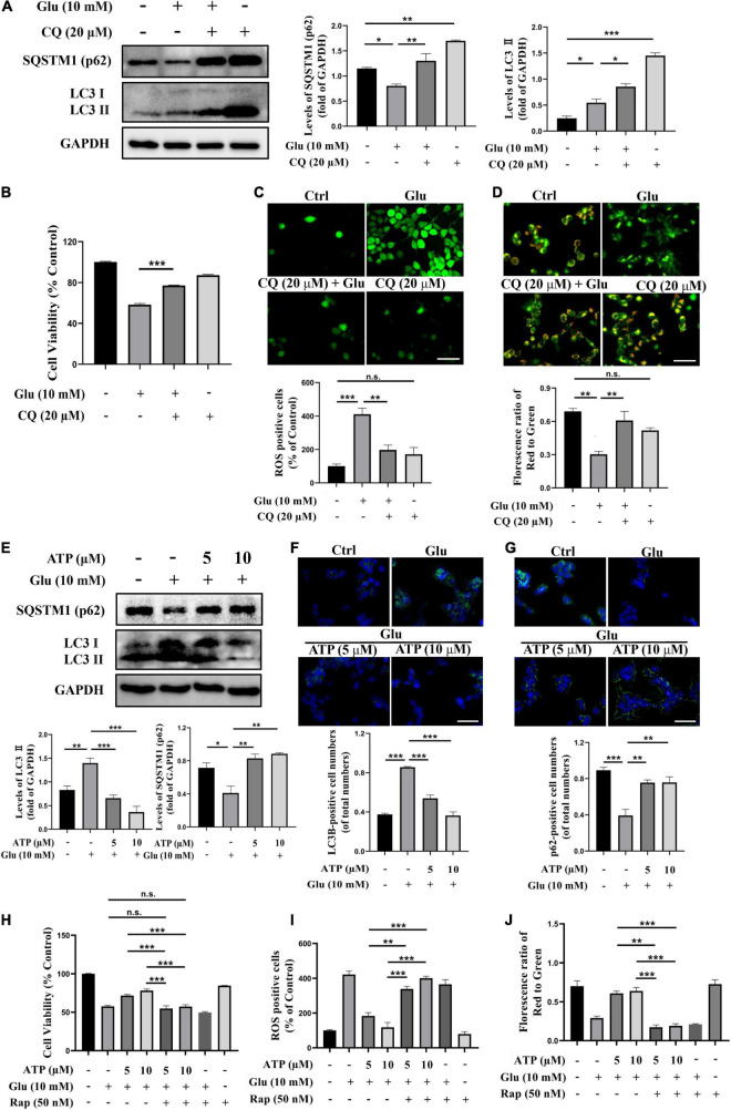
FIGURE 3.
Extracellular ATP prevents glutamate-induced excitotoxicity via suppressing autophagy in SH-SY5Y cells. (A) The protein expression of LC3 and p62 were determined by western blot in SH-SY5Y cells treated with 10-mM Glu in the presence or absence of chloroquine (CQ) (20 μM), and expression of GAPDH was served as loading control. The quantitation of LC3 and p62 expressions were calibrated. (B) Cell viability was evaluated. (C) Treated SH-SY5Y cells were stained with DCFH-DA, and the changes were shown in histograms as percentage with control. Scale bar, 100 μm under 20× magnification. (D) Treated SH-SY5Y cells were stained using JC-1, and the changes were shown in histograms as percentage with control. Scale bar, 100 μm under 20× magnification. (E) The protein expressions of LC3 and p62 were determined by western blot in SH-SY5Y cells treated with 10-mM Glu in the presence of ATP (5 and 10 μM), and expression of GAPDH was served as loading control. The quantitation of LC3II and p62 expressions was calibrated. (F) LC3II and (G) QSTM1/p62 detected using immunocytochemistry were photographed under fluorescent microscope, and images were analyzed. (H) Cell viability was detected. (I) ROS-positive cells were calibrated. (J) Treated SH-SY5Y cells were stained using JC-1, and the changes were presented. All data in bar charts present mean ± SEM from at least three independent experiments. *P < 0.05, **P < 0.01, ***P < 0.001 versus the ATP treated group or the glutamate group. n.s., no significance.

