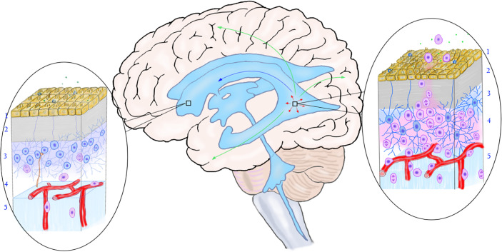
Figure 3.
The relationship between the mutation of neural stem cells in the subependymal zone and the occurrence of multicentric glioma. The rectangular box on the left shows the normal subependymal area that is not affected by the tumor, and the oval area on the left shows the normal microstructure of the subependymal area. From the ventricle to the brain parenchyma, there are ependymal layer (layer 1), gap zone (layer 2), astrocyte zone (layer 3), loose layer (layer 4), and brain parenchyma (layer 5). It can be seen that neural stem cells differentiate into less primary progenitor cells, and the local cell hierarchy is regular. The rectangular frame on the right shows the location of the mutant neural stem cells, and the oval area on the right shows the microstructure of the area. As shown in the figure, early gene mutations occurred in neural stem cells in this area, leading to active cell proliferation and differentiation into progenitor cells. The primary progenitor cells migrated along the white matter fibers and blood vessels into the brain parenchyma, replicated, differentiated, and accumulated mutations to form tumors. The arrow shows the migration pathway of the mutant progenitor cells: the green arrow shows the migration into the brain parenchyma, the red arrow shows the local migration along the subependymal zone, and the blue arrow shows the migration along the cerebrospinal fluid (to be verified). (Yellow cells in layer 1, ependymal cells; blue cells in layer 2, neural stem cells; pink cells in layer 3, progenitor cells; brown-yellow cords in layer 3 and layer 4, axons and synapses of distant neurons; green dots, chemical transmitters in cerebrospinal fluid).

