
Figure 2.
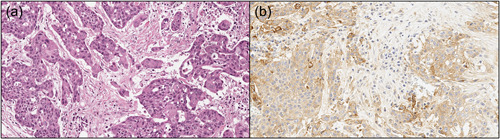
A classic example of a case of invasive oral squamous cell carcinoma, poorly differentiated, showing diffuse positivity for PD‐L1. (a) Hematoxylin & eosin stain, original magnification ×20; (b) PD‐L1 immunostaining with 22C3 antibody, 90% TPS, 90 CPS. CPS, combined positive score; PD‐L1, programmed cell death‐ligand 1; TPS, tumor proportion score.