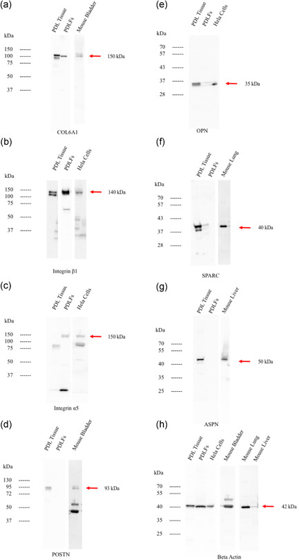
Figure 7.

The western blot analysis for collagen type6A1 (a), integrin β1 (b), integrin α5 (c), POSTN (d), OPN (e), SPARC (f), ASPN (g), and beta‐actin (h). Protein expression showed that collagen type6A1 and integrin β1 was highly expressed in both PDL tissue and PDLFs. OPN, POTN, ASPN, and SPARC were highly expressed in PDL tissue while integrin α5 was highly expressed in PDLFs. As positive controls, the extracts from mouse lung, mouse bladder, mouse liver, and Hela cells were utilized in each blotting separately. PDL, periodontal ligament; PDLF, PDL fibroblast
