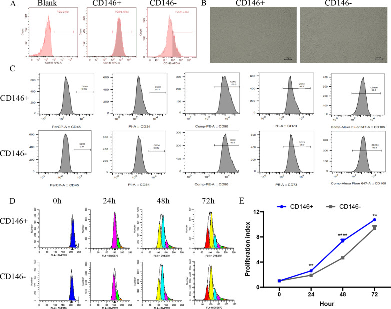
Fig. 1.
Comparison of biological characteristics of CD146 +/− MSCs subpopulations. A Flow Cytometry detection for CD146 expression in UC-MSCs after separation using magnetic beads. B The cellular morphology of CD146 +/− subsets after separation using magnetic beads. C Flow Cytometry detection of surface markers of CD146 +/− MSCs subsets: CD105, CD73, and CD90 were positive, and CD45 and CD34 were negative. D CD146 +/− MSCs subsets were labeled with Dye eFluor™ 670, and cell proliferation was assayed by flow cytometry at 0 h, 24 h, 48 h and 72 h. E Cell proliferation index calculated based on flow experimental results

