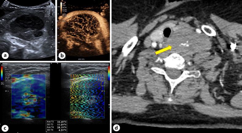
Fig. 1.
US: left side hypoechoic, irregular, inhomogeneous neck mass (5 × 53 × 25.0 cm) with multiple central calcifications (a), CEUS: homogeneous trabecular early hyperenhancement (b), moderate elasticity on SWE 28–33 kPa (c). CT: mass with anterolateral dislocation of the common carotid artery in the axial plane (d). SWE, shear wave elastography.

