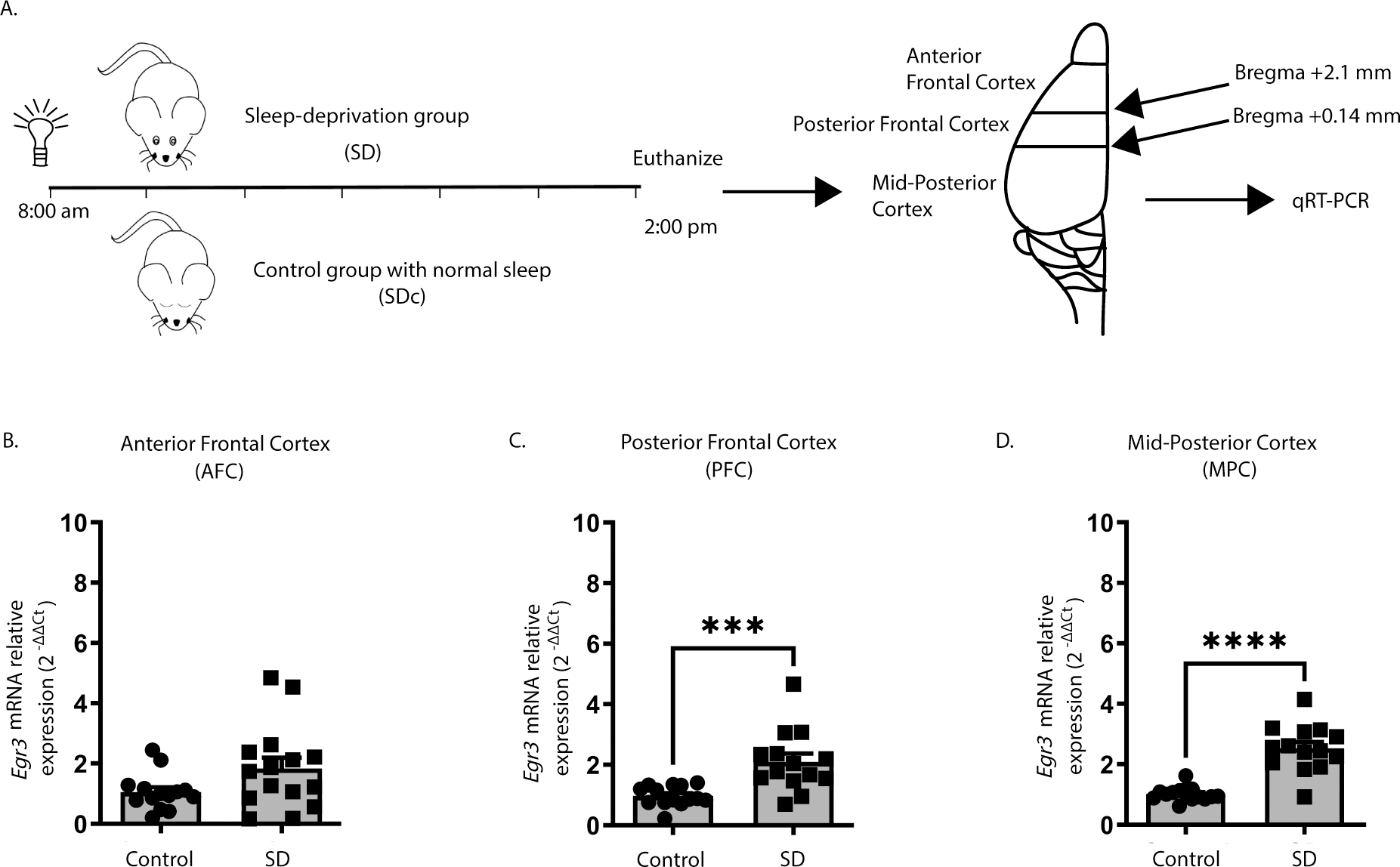
Figure 1. Sleep deprivation upregulates Egr3 in a region-dependent manner in the frontal cortex.

(A) SD protocol. In WT mice quantitative RT-PCR shows that 6h of SD (B) does not increase Egr3 expression in AFC regions (t27 = 1.956; p = 0.0609; SDc, n = 14; SD, n = 15 ), but significantly upregulates Egr3 mRNA in (C) PFC (t26 = 3.979, p = 0.0005; SDc, n = 14; SD, n = 14) and (D) MPC (t27 = 7.307; p < 0.0001; SDc, n = 15; SD, n = 14) regions, compared to SDc. Unpaired student’s t-test, *** p < 0.001, **** p < 0.0001. Values represent means ± SEM. (Abbreviations: AFC- anterior frontal cortex; PFC- posterior frontal cortex; MPC- mid to posterior cortex; SD- sleep deprivation; SDc- SD control; WT- wildtype; h: hours).
