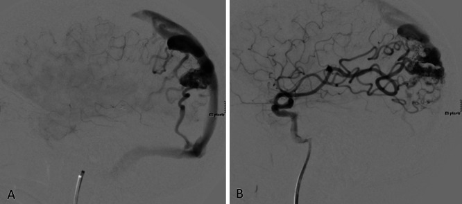
FIG. 1.

Cerebral angiography of left parietal AVM pre- and postembolization. A: Preembolization angiogram of the left internal carotid artery shows a lateral view of the malformation draining into the superior sagittal sinus and left transverse sinus. B: Postembolization angiogram of the left internal carotid artery demonstrates obliteration of the feeding MCA branch.
