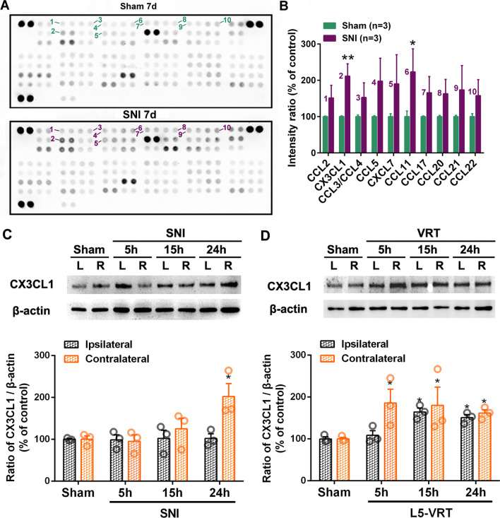
Fig. 3.
Comparison of L5-VRT and SNI-induced expression of CX3CL1 in bilateral ACC. Representative protein microarray results (A) and the quantification analysis (B) showing that SNI triggered upregulations of chemokines CX3CL1 and CCL11 in contralateral ACC.*p < 0.05, **p < 0.01 versus sham group (n = 3 rats/group, one-way ANOVA). C, D Representative western blotting of CX3CL1 protein levels in bilateral ACC following SNI (C) and L5-VRT (D) are shown on the top and the quantification results are shown below. Significant differences are observed on the contralateral side in the SNI group but on the bilateral side in the L5-VRT group. *p < 0.05 versus sham group (n = 3 rats/group, one-way ANOVA)

