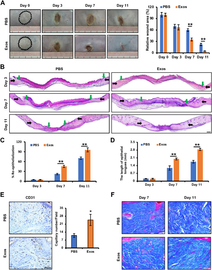
Fig. 2.
iPSCs-KCs-Exos accelerate deep second-degree burn wound healing in vivo. A Representative images of wounds treated with PBS or iPSCs-KCs-Exos on days 0, 3, 7, and 11 after burning and quantitative analysis of wound area in each group. n = 6 mice per group. B H&E staining of wounded skin sections treated with PBS or iPSCs-KCs-Exos on days 3, 7, and 11 after burning. Black arrows indicate the dermal border; green arrows indicate the epidermal margin. Scale bar, 400 μm. C Quantitative analysis of the re-epithelialization ratio of wounds in each group. D Quantitative analysis of the length of epithelial tongues of wounds in each group. E Immunohistochemistry staining for CD31 wounds and quantitative analysis of the numbers of stained capillaries in each group on days 11 after burning. Scale bar, 50 μm. F Masson’s trichrome staining of wounds in each group on days 7 and 11 after burning. Scale bar, 50 μm. *P < 0.05, **P < 0.01

