Fig. 9.
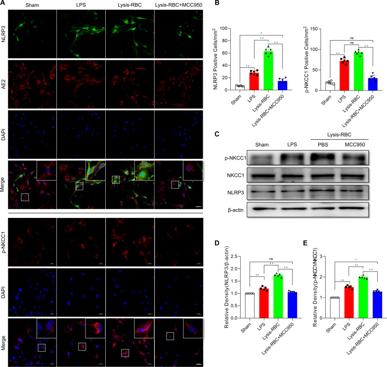
NLRP3/p-NKCC1 pathway activation in primary choroid plexus epithelial cells after LPS or lysis-RBC stimulation. A Immunoreactivity of NLRP3 (green) and p-NKCC1 (red, down) as shown by immunofluorescence microscopy in rat primary choroid plexus epithelial cells exposed to LPS or lysis-RBCs and treated with MCC950. Primary choroid plexus epithelial cells were identified and stained with AE2. Nuclei were stained with DAPI. Bar = 50 μm. B Morphometric analysis of NLRP3 and p-NKCC1 immunoreactivity in primary choroid plexus epithelial cells exposed to LPS or lysis-RBCs and treated with MCC950 (n = 6, one-way ANOVA). C–E Western blot quantitative analysis of NLRP3 and p-NKCC1 in primary choroid plexus epithelial cells after different treatments (n = 5; one-way ANOVA). The values are expressed as the means ± SDs; **p < 0.01 and *p < 0.05, ns, p > 0.05

