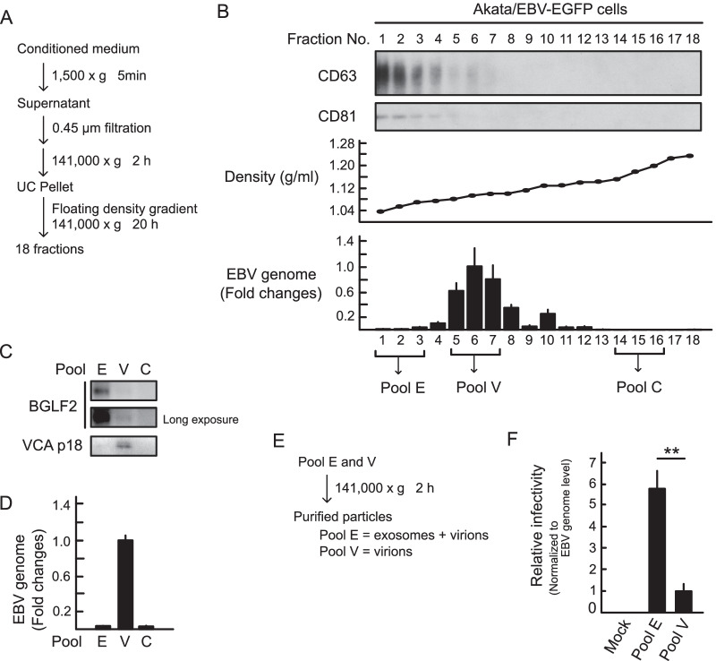
Fig. 4.
BGLF2-containing exosomes are released by infected cells. A Centrifugation protocol and workflow for separation and enrichment of exosomes and virions. B Exosome marker proteins were enriched in fractions #1-3 after iodixanol floating density gradient centrifugation. For immunoblotting, proteins in each fraction were concentrated by acetone precipitation. CD63 and CD81 served as exosome markers. The EBV genome in each fraction was quantified by qPCR. C and D Pool E (fractions #1-3) containing the BGLF2 protein. Each pool (E, fractions #1-3; V, fractions #5-6; and C, fractions #14-16) was analyzed by immunoblotting (C) and qPCR (D). Data are presented as the mean ± SE. Viral capsid antigen (VCA) p18 served as a virion marker. E Workflow for particle purification. F Akata(-) cells were incubated with purified particles for 24 h. GFP positivity was determined by FACS. The EBV genome of each pool was quantified by qPCR. The results are presented as the mean ± SE of four independent experiments after normalization to the EBV genome levels and as relative infectivity to pool V (infectivity value of 1). Double asterisks, p < 0.01

