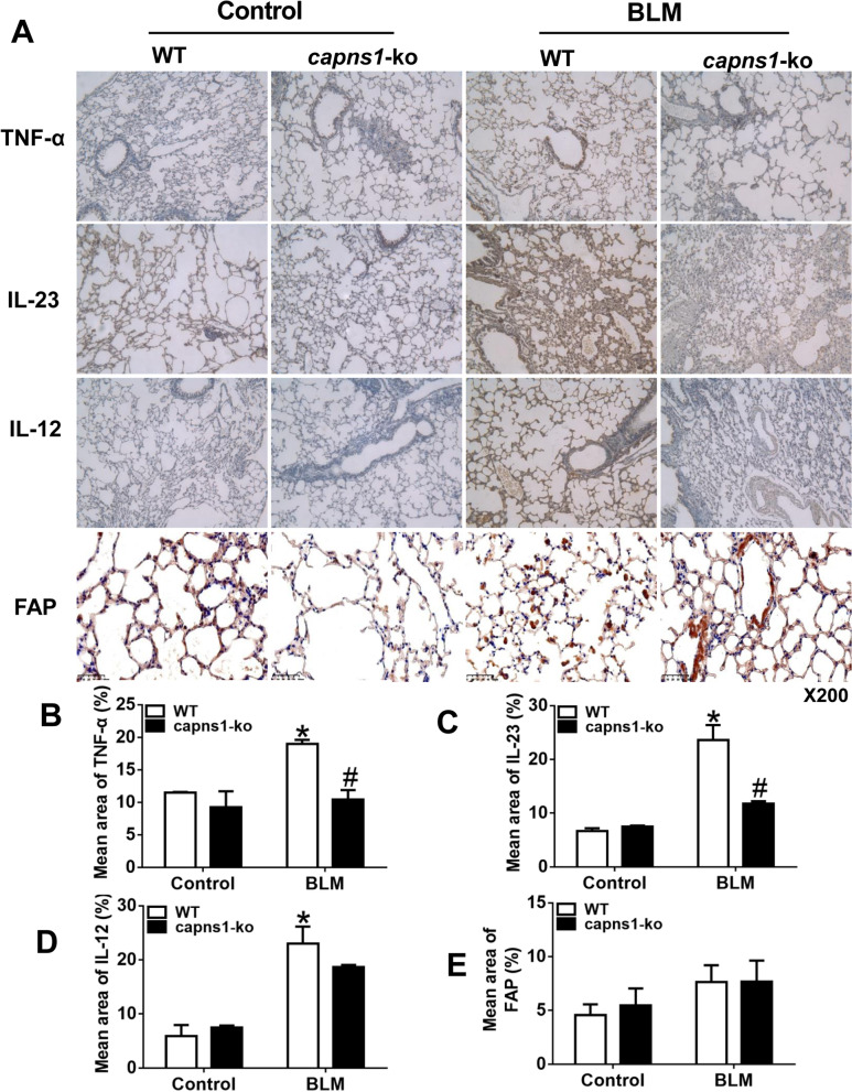
Fig. 7.
Immunohistochemical analysis of TNFα, IL-12 and IL-23 in lung tissues of the bleomycin model of SSc mice. A Lung sections of sham treated (Control) or BLM treated (bleomycin model of SSc) WT or Capns1-ko mice were stained for TNFα, IL-23, IL-12 or FAP. Scale bars, 50 μm, magnification X200. (B, C, D,E) Mean staining area were quantified for TNFα B, IL-23 C, IL-12 D or FAP. E. The test of Homogeneity of variance showed P < 0.1, nonparametric test was applied. Data are mean ± SD. n = 10, *: P < 0.05, vs. sham treated WT mice; #: P < 0.05, vs. bleomycin model of SSc WT mice. OD: optical density

