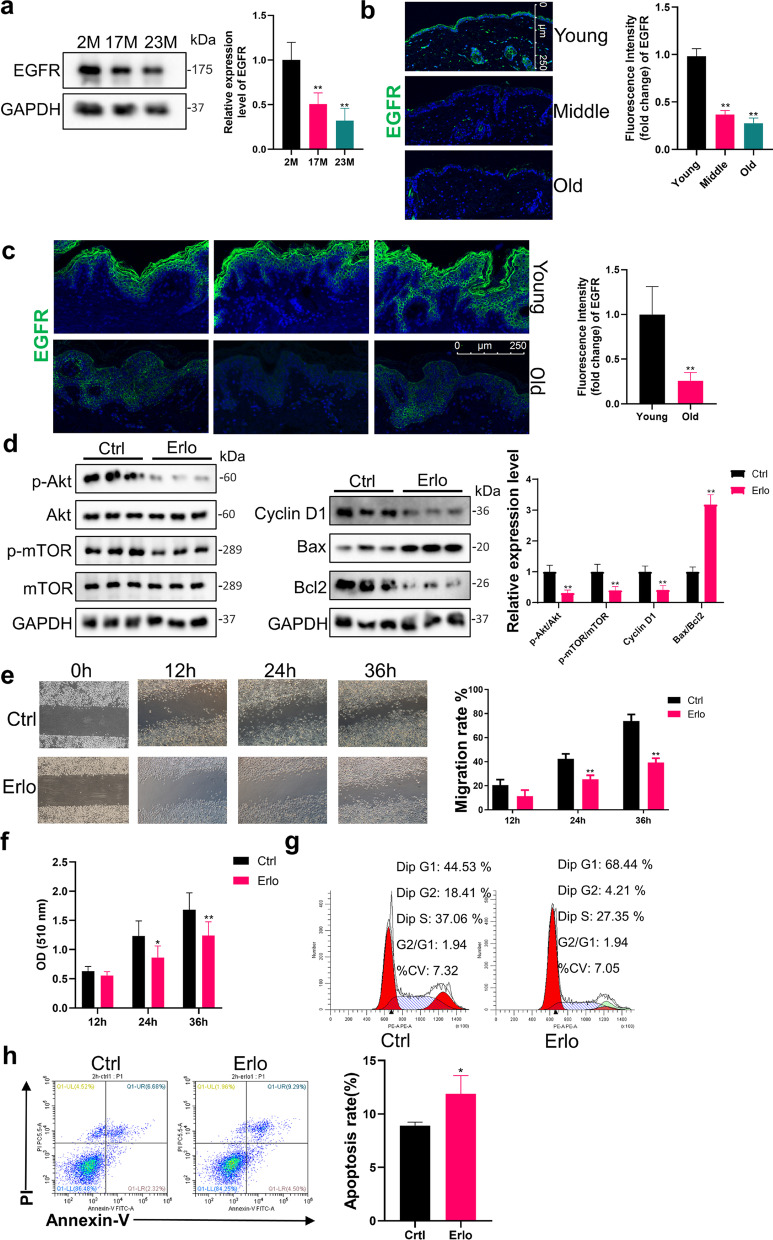
Fig. 2.
Declines in EGFR expression restricted the activation of PI3K/Akt in EpiSCs. a Western Blot was used to measure the expression level of EGFR in epidermis from the back of 2 M, 17 M and 23 M rats. N = 6. *P < 0.05, **P < 0.01 versus the 2 M group. b Immunofluorescence images of young, middle and aged mice skin labeled with antibodies against EGFR [secondary antibodies are color-coded as shown]. Sections were co-stained with DAPI (blue) to visualize nuclei. N = 6. Scale bar, 250 μm. *P < 0.05, **P < 0.01 versus the young group. c Immunofluorescence images of young and old human skin labeled with antibodies against EGFR [secondary antibodies are color-coded as shown]. Sections were co-stained with DAPI (blue) to visualize nuclei. N = 3. Scale bar, 250 μm. *P < 0.05, **P < 0.01 vs. the young group. d EpiSCs were randomly divided into two groups that incubated with PBS or 2 μM Erlotinib dissolved in Epilife medium for 24 h. Western Blot was performed. N = 3 for each group. *P < 0.05, **P < 0.01 vs. the control group. e Confluent EpiSCs were randomly divided into two groups that received PBS or 2 μM Erlotinib dissolved in Epilife medium and subjected to in vitro scratch-wound assays. N = 3 for each group. *P < 0.05, **P < 0.01 versus the control group at time-point. f SRB assay was used to evaluate the proliferation ability of EpiSCs treated with PBS or 2 μM Erlotinib. N = 6 for each group. *P < 0.05, **P < 0.01 vs. the control group at time-point. g and h Flow cytometry was further used to quantify cell cycle distribution and apoptosis in cells treated with PBS or 2 μM Erlotinib for 24 h. *P < 0.05, **P < 0.01 vs. the control group. N = 3 for each group

