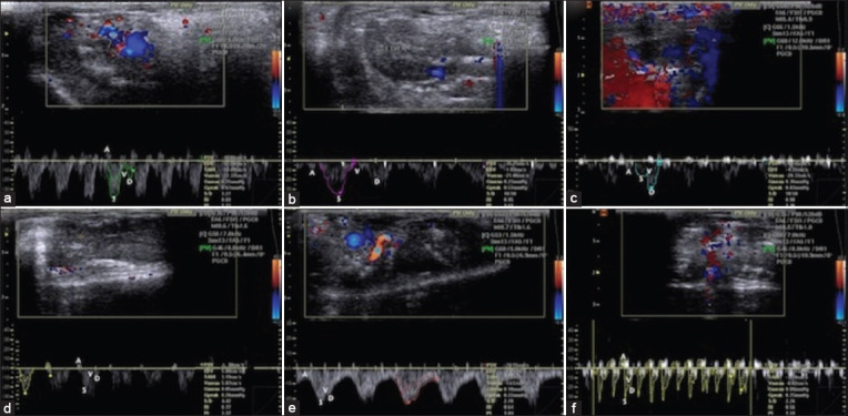
Figure-4.

Color and spectral Doppler USG of hepatic venous flow direction and waveform in healthy, acute aflatoxicosis rat model, and other different treated groups. (a) Hepatic venous flow direction and waveform images in healthy control rats; the hepatic venous waveform was phasic, and the direction of flow was predominantly antegrade. (b) By contrast, in the acute aflatoxicosis rat model, the hepatic venous waveform was phasic, and the direction of flow was antegrade corresponding to the three waves (A, S, and D). Abnormally mild decreased phasicity was observed in the hepatic vein of the acute aflatoxicosis rat model. (c) In the 4th week after IV BM-MSCs injection of rats, the hepatic venous waveform showed phasicity and the direction of blood flow was antegrade that corresponding to the three waves (A, S, and D) through color and spectral Doppler USG. (d) Conversely, in the 4th week after IH MSCs injection of rats, normal hepatic venous flow direction and waveform were observed: The hepatic venous waveform was phasic and predominantly antegrade with a tetrainflectional waveform (A, S, V, and D waves) that was mostly below the baseline in spectral Doppler USG. (e) The IV HDCs injected rats showed a phasic waveform with an antegrade direction of hepatic venous flow, which corresponded to the three waves (A, S, and D) through color and spectral Doppler USG, at 4 weeks after injection. (f) By contrast, normal hepatic venous flow direction and waveform were observed via color and spectral Doppler USG at 4 weeks after injection in the IH HDCs injected rats: The hepatic venous waveform was phasic and predominantly antegrade with tetrainflectional waveform. IV=Intravenous, IH=Intrahepatic, BM-MSCs=Bone marrow mesenchymal stem cells, HDCs=Hepatogenic differentiated cells, USG=Ultrasonography.
