
Figure 3.
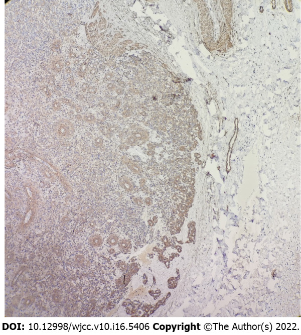
Immunohistochemical staining of smooth muscle actin showed that tumor cells and surrounding vascular wall were positive. (Smooth muscle actin 4 ×).
Official websites use .gov
A
.gov website belongs to an official
government organization in the United States.
Secure .gov websites use HTTPS
A lock (
) or https:// means you've safely
connected to the .gov website. Share sensitive
information only on official, secure websites.
Immunohistochemical staining of smooth muscle actin showed that tumor cells and surrounding vascular wall were positive. (Smooth muscle actin 4 ×).