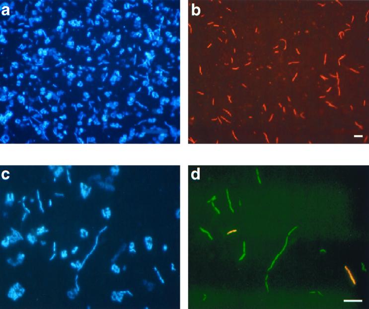
FIG. 2.
Identification by FISH of Bacteria in samples from crystallizer ponds. Identical microscopic fields were visualized with an epifluorescence microscope using filter sets specific for DAPI (a and c) and the fluorochromes used for probe labeling (b and d). (a and b) Cells from the crystallizer in Majorca hybridized with Cy3-labeled probe EHB412. (c and d) Cells from CR-30 (Alicante) hybridized with fluorescein-labeled probe EHB586 and Cy3-labeled probe EHB1451. Bar, 5 μm.

