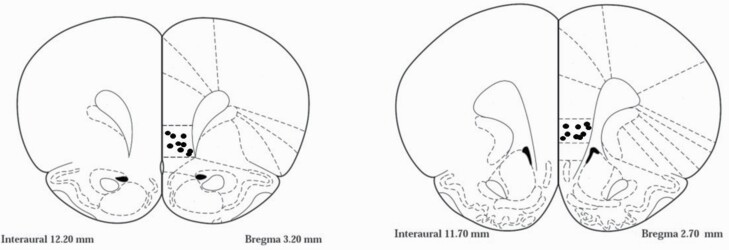
Figure 1.
Schematic drawing illustrating cannulae placements representation of a sample of animals in the infralimbic cortex on coronal view at position +3.2 mm and +2.7 mm anterior to bregma (according to Paxinos and Watson, 1998). Black dots indicate the locations of the tips of the cannula.

