
Figure 2.
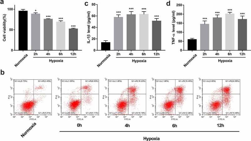
Establishment of an in vitro hypoxia/reoxygenation injury model in BEAS-2B cells. (a-d) BEAS-2B cells were exposed to hypoxic conditions for 0, 2, 4, 6, or 12 h, and then maintained in normoxic conditions (reoxygenation) for the same treatment time-period with hypoxia. (a) Cell viability was measured by CCK-8 assay. (b) Cell apoptotic rate was measured using flow cytometry. (c and d) The secretion levels of IL-1β and TNF-α were examined by ELISA assay.