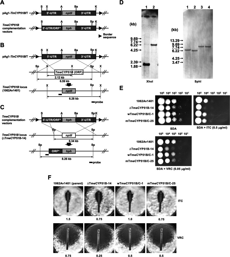
FIG 2.
Disruption of the CYP51B homolog (TmeCYP51B) of T. mentagrophytes 1062Av1401 and subsequent reintroduction of the wild-type and mutated copy of TmeCYP51B by a gene replacement strategy. (A) Schematic representation of a binary TinCYP51B-targeting vector, pAg1-TinCYP51B/T. The nptII cassette is composed of Aspergillus nidulans trpC promoter (PtrpC), E. coli neomycin phosphotransferase gene (nptII), and A. fumigatus cgrA terminator (TcgrA). Border sequences are the specific regions that delineate the DNA to be transferred during Agrobacterium tumefaciens-mediated transformation. Restriction enzyme site: A, ApaI; Ba, BamHI; E, EcoRI; K, KpnI; P, PstI; S, SpeI; X, XhoI. (B) Schematic representation of the TmeCYP51B locus before and after homologous recombination. (C) Schematic representation of the TmeCYP51B locus in the ΔTmeCYP51B-14 before and after complementation by two kinds of binary vectors pAg1-wTmeCYP51B/C and pAg1-mTmeCYP51B/C. DNA fragments (TmeCYP51B1 and TmeCYP51B2) containing the 5′ UTR of the TmeCYP51B gene and the ORFs encoding wild-type or mutated TmeCYP51B proteins (ORF*) were subcloned into the pAg1-TinCYP51B/T upstream (SpeI/ApaI) of the hph cassette, respectively (Table 2). (D) Southern blotting. Aliquots of approximately 10 μg of genomic DNA from each strain were digested with XhoI or SphI and separated by electrophoresis on 0.8% (wt/vol) agarose gels. Lane 1, 1062Av1401 (parent strain); lane 2, ΔTmeCYP51B-14 (TmeCYP51B disruptant); lane 3, wTmeCYP51B/C-1 (revertant harboring the wild-type TmeCYP51B gene); lane 4, mTmeCYP51B/C-25 (mutant harboring the mutated TmeCYP51B gene). A fragment of about 480 bp of the TmeCYP51B locus was amplified by PCR with a pair of the primers P57 and P36 (Table S1) and used as a hybridization probe. DNA standard fragment sizes are shown on the left. (E and F) Evaluation of ITC and VRC susceptibility in the four T. mentagrophytes strains (1062Av1401, ΔTmeCYP51B-14, wTmeCYP51B/C-1, and mTmeCYP51B/C-25) by serial dilution drug susceptibility assays (E) and Etest assays (F). For serial dilution drug susceptibility assays, spores from each strain were spotted at different dilutions on SDA plates, as described in Materials and Methods. The plates were incubated at 28°C for 3 to 5 days (serial dilution drug susceptibility assays) or 4 days (Etest assays).

