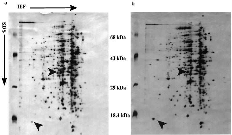
FIG. 1.
Silver-stained 2-D SDS-PAGE of total cellular proteins extracted from 16-h cultures of L. monocytogenes B73 (a) and L. monocytogenes B73-MR1 (b) grown at 30°C, which were firstly separated by isoelectric focusing (IEF) (1.6% [vol/vol] Ampholine [Pharmacia] [pH 5 to 7] and 0.4% [vol/vol] Ampholine [Pharmacia] [pH 3.5 to 9.5]) in the horizontal direction, followed by SDS-PAGE (16.5% acrylamide-bisacrylamide [44:0.8]) in the vertical direction. Directions of isoelectric focusing and SDS-PAGE are indicated by arrows. Protein differences are indicated by arrow heads.

