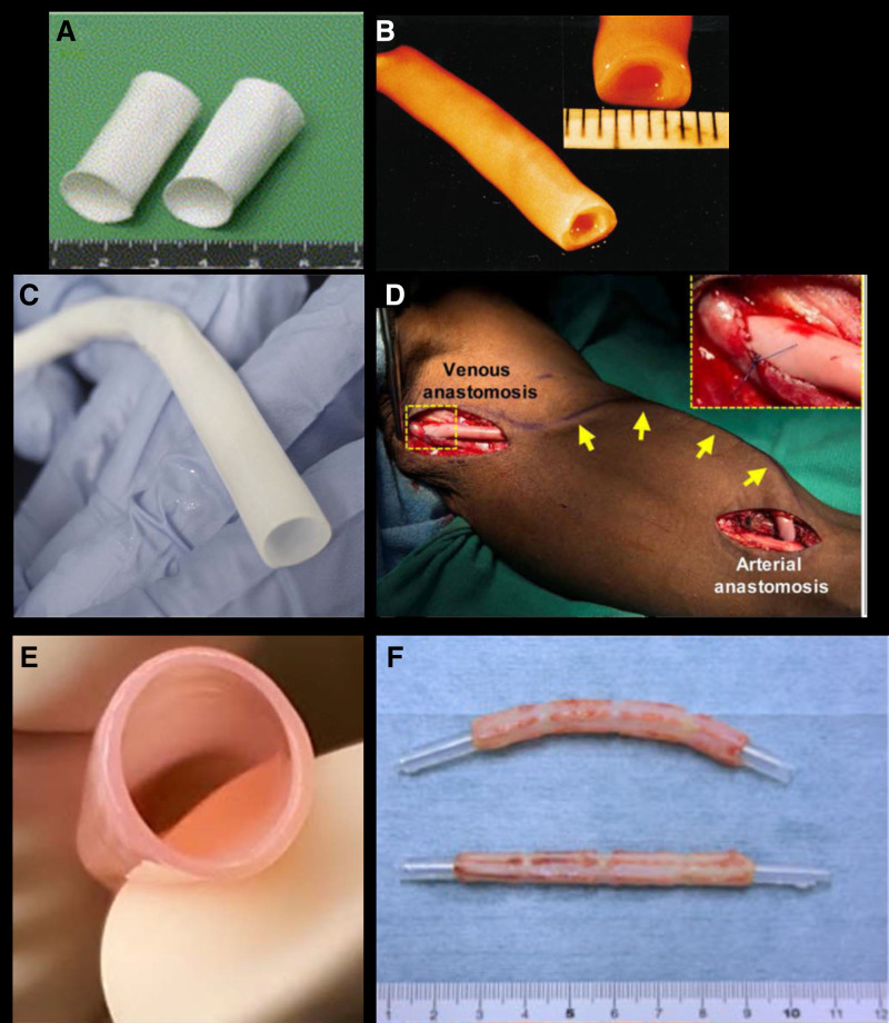
Figure 4.
Presentation of various approaches to tissue engineered vascular grafts. A, Macroscopic image of polymeric scaffold before cell seeding. B, Gross image of biological tissue-engineered vascular graft (TEVG) produced by cell sheets rolled around a mandrel. C, A HAV ready to store/implant after decellularization.115 D, Intraoperative image of HAV implanted in the arm of a patient with end-stage renal disease as an arteriovenous conduit for hemodialysis.133 E, TEVGs produced from fibroblast-populated tubular fibrin constructs, matured in pulsatile bioreactor, and decellularized. F, Biotube made of fibrous tissue formed by foreign body reaction to the implanted mandrel. A, Reproduced from Shin’oka et al161 with permission. Copyright ©. B, Reproduced from L’Heureux et al128 with permission. Copyright ©. E, Reproduced from Syedain et al137,140 with permission. Copyright ©. F, Reproduced from Nakayama et al143 with permission. Copyright ©.

