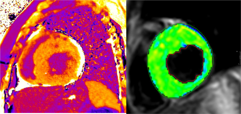
Figure 44.
On the left is an extracellular volume map showing globally elevated elevated extracellular volume fraction (ECV) in a 14 year old patient with hypertrophic cardiomyopathy (HCM). On the right is a T2 map of left ventricular myocardium with T2 value of 69 ms ± 1.2 also in a 14 year old patient with HCM.

