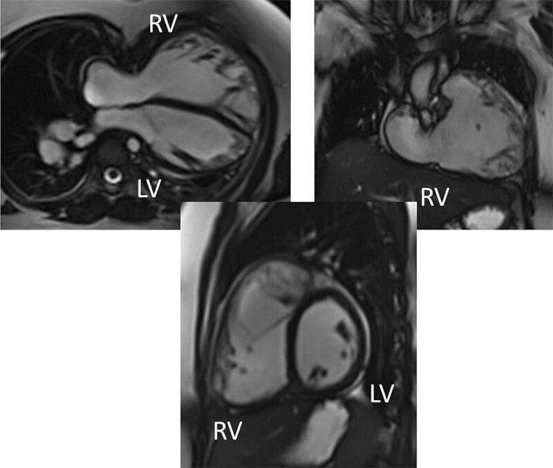
Figure 7.
Ventricular function and volumes in tetralogy of Fallot. The 4-chamber (upper left), right ventricle (RV) two chamber (upper right) and short axis (lower panel) views of the patient in Figure 6 with volume overload of the RV. LV indicates left ventricle.

