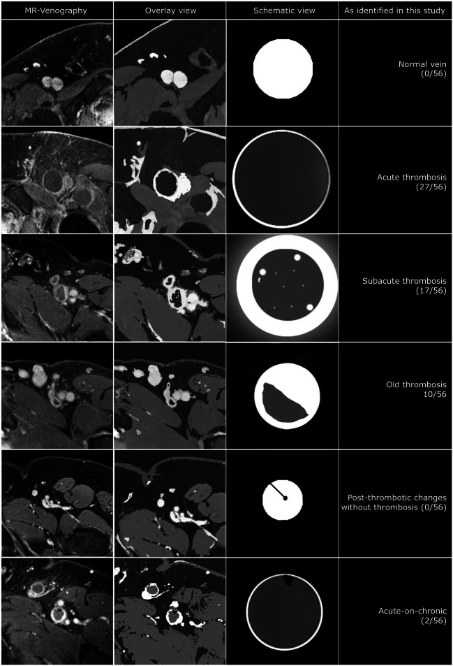
Fig. 1.
Thrombus characteristics identified using MR-venography. Normal vein: homogeneously opacified hyperintense vein lumen. No luminal defect or perivascular) wall changes. Acute thrombosed vein: dilated homogeneously hypointense vein lumen with small enhancing rim of contrast depicting the vein wall. No (perivascular) wall changes (no halo sign). Subacute thrombosed vein: Still dilated low intensity vein lumen with thick enhancing rim of contrast, part vein wall thickening and part perivascular edema (halo sign). There are some small hyperintense areas within the thrombus as sign of recanalization. Old thrombosed vein: the vein lumen is reduced to a more ‘normal’ vein size with an opacified part (open lumen/vein wall) and a low intensity part that is still filled with thrombus-like tissue. Post-thrombotic vein: the vein lumen is smaller than the normal vein and homogeneously opacified except for 1 or more sharply demarcated very low intensity black dots and/or lines adhering to the vein wall. This represents (fibrotic) scar tissue (post-thrombotic venous scarring). Acute-on-chronic thrombosed vein: as in an acute deep vein thrombosis there is a dilated lumen with mostly hypointense material but additionally there are signs of a previous thrombotic event that has left scar tissue markings (very hypointense dots and lines)

