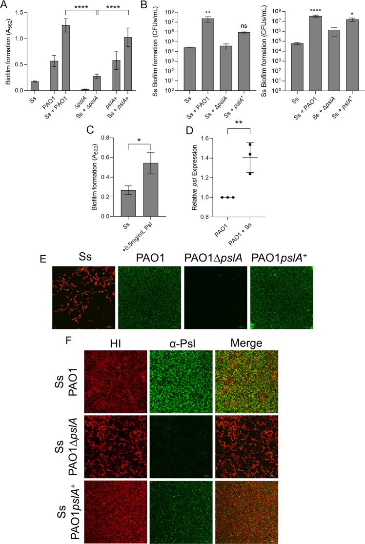
Fig. 3. P. aeruginosa exopolysaccharide Psl promotes S. salivarius biofilm formation.
Ss was co-cultured with P. aeruginosa PAO1 strains (A) PAO1∆pslA and PAO1 pslA+ in TSBYE medium with 1% sucrose in a 96-well plate for 16 h at 37 °C with 5% CO2 (n = 3 biological replicates, 3 technical). Biofilm biomass was then measured using crystal violet staining. One-way ANOVA with Dunnett’s multiple comparisons test. B Quantification of Ss biofilm-forming cells after co-culturing with PAO1, PAO1∆pslA, and PAO1 pslA+ in TSBYE (left) and SCFM2 [50] in a 6-h, 6-well model at 37 °C with 5% CO2 (n = 3 biological replicates, each with 3 technical replicates). One-way ANOVA with Šίdák’s multiple comparisons test. C 0.5 mg/mL purified Psl was added to Ss single cultures in TSBYE with 1% sucrose in a 96-well 16-h biofilm. Crystal violet staining was used to quantify biofilm biomass. D qPCR quantification of P. aeruginosa pslA expression compared to 16S rRNA control. Student’s t test. Fluorescence microscopy images at 60× magnification of 16-h single (E) and dual species (F) biofilms of Ss and PAO1, PAO1∆pslA, and PAO1pslA+ cultured in TSBYE supplemented with 1% sucrose. Ss was stained with hexidium iodide, and Psl was stained with a FITC-conjugated α-Psl monoclonal antibody. Scale bar: 20 μm. *p < 0.05, **p < 0.01, ****p < 0.0001.

