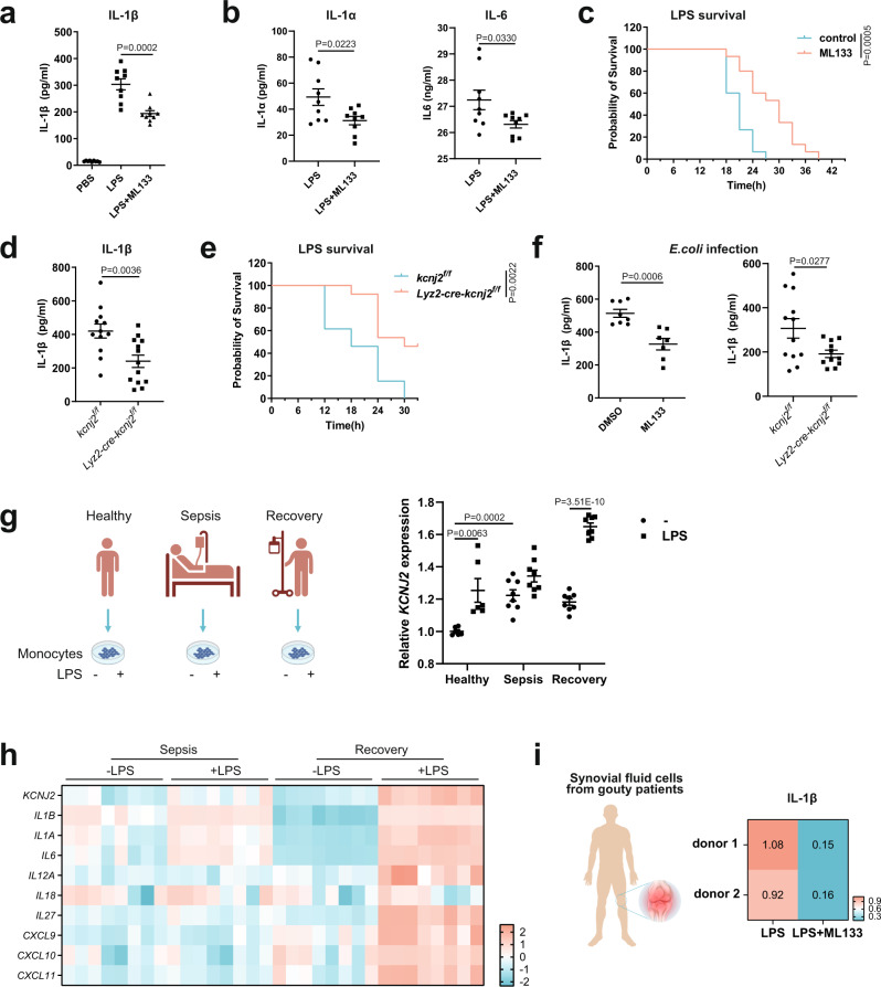
Fig. 7. Kir2.1 repression alleviates inflammation triggered by pathogenic or danger signals in mouse models and human samples.
a IL-1β levels in serum from septic mice measured by ELISA. (n = 9; mean ± SEM) (b) IL-1α and IL-6 levels in serum from septic mice measured by ELISA. (n = 9, mean ± SEM) (c) Survival rates of sepsis model mice (n = 15, log-rank test [Mantel–Cox]). d IL-1β levels in serum from septic mice measured by ELISA (Kcnj2f/f, n = 12; Lyz2-cre-Kcnj2f/f, n = 13; mean ± SEM). e Survival rates of sepsis model mice (n = 13; log-rank test [Mantel–Cox]). f IL-1β levels in serum from septic mice measured by ELISA (DMSO, n = 8; ML133, n = 7; Kcnj2f/f, n = 12, Lyz2-cre-Kcnj2f/f, n = 11; mean ± SEM). g The expression of KCNJ2 in monocytes from healthy donors, sepsis patients, and those who recovered from sepsis with or without LPS63. Data sourced from the GEO database (GSE46955). (n = 6, 8, 8 respectively; mean ± SEM). h Heatmap showing the expression of Kir2.1 and inflammatory genes in monocytes from septic patients and those who recovered from sepsis in response to LPS63. Data sourced from the GEO database (GSE46955). (n = 8, 8 respectively; mean ± SEM). i Relative IL-1β levels in the supernatant of cultured synovial fluid cells from two gouty patients analyzed by ELISA. Two-tailed unpaired Student’s t-test. Source data are provided as a Source Data files.

