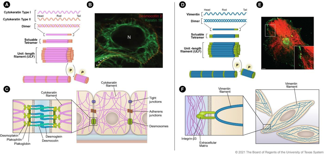
Figure 1. Filamentous assembly and localization of cytokeratin and vimentin:
A) Cytokeratin assembly initiates with type I and type II heterodimer formation. The heterodimers combine to form soluble tetramers that interact to form ULFs made up of eight monomers. ULFs are then assembled in a phosphorylation-dependent manner into 10-nm filaments. B) Survey fluorescence micrograph of MDCK cells that express YFP-labeled desmocollin (red) and mRFP-labeled human cytokeratin 18 (green). Image reprinted with permission from Quinlan et al., J. Cell Sci., 130, 3437–3445 (2017). C) Schematic of cytokeratin distribution in an epithelial cell with cell-cell adhesion processes. Zoomed inset shows the protein-protein interactions between cytokeratin filaments and desmosomes. D) Vimentin assembles as homodimers that combine to form soluble tetramers and then ULFs made up of eight monomers. ULFs are then assembled in a phosphorylation-dependent manner into filaments. E) Transformed human bone marrow endothelial cells stained for integrin β3 (green) and vimentin (red). Image reprinted with permission from Bhattacharya et al., J. Cell Sci., 122, 1390–1400 (2009). F) Schematic of vimentin distribution in a mesenchymal cell. Zoomed inset indicates the vimentin filament interaction with β3 integrin that anchors the filament to the extracellular matrix.

