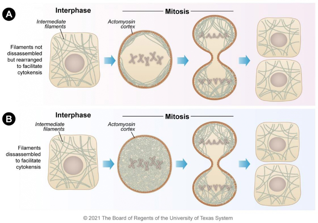
Figure 2. Localization mechanisms for vimentin and cytokeratin during mitosis.
Both vimentin and cytokeratin filaments exist as filamentous forms during interphase. During mitosis, the filaments either A) remain filamentous but are organized to the actin cortex to clear the mitotic plate or B) become disassembled into unit-length filaments to clear the mitotic plate. Both mechanisms enable the efficient completion of cytokinesis resulting in daughter cells.

