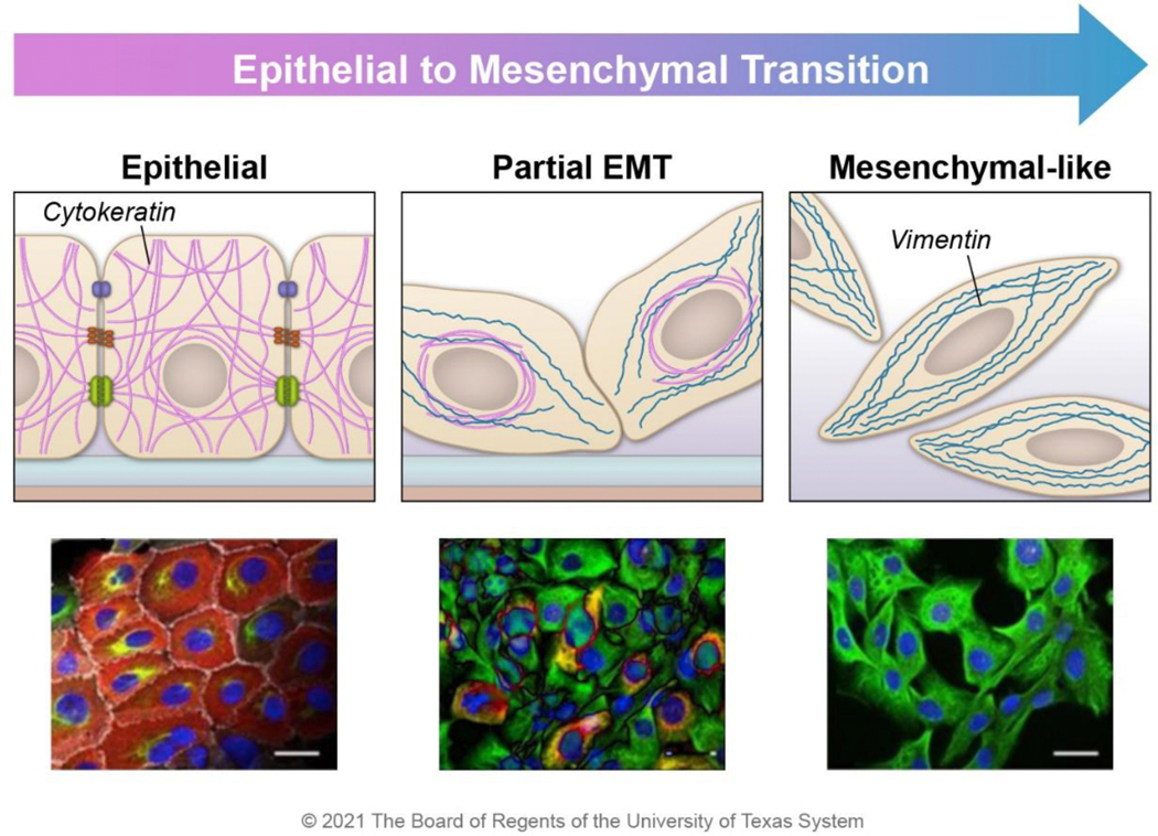
Figure 4. Intermediate filament expression changes during EMT.
Epithelial-like cancer cells express cytokeratin (red), which aids in stress resistance and supports cell-cell adhesion; these cells do not express vimentin. As cells progress through EMT, mesenchymal markers such as vimentin (green) begin to be expressed. In E/M hybrid cells, cytokeratin is perinuclearly localized, and the vimentin filament network stretches from the nucleus to the periphery of the cell. At the far end of the EMT spectrum, epithelial markers are lost. The cartoons were constructed based on the observed morphology of HMLER cells during the transition from epithelial (CD104+ CD44low) to hybrid E/M (CD104+ CD44hi) to mesenchymal (CD104− CD44hi) populations 13. Images are reprinted with permission from Kröger, C. et al. Acquisition of a hybrid E/M state is essential for tumorigenicity of basal breast cancer cells. Proc. Natl. Acad. Sci. 116, 7353–7362 (2019).

