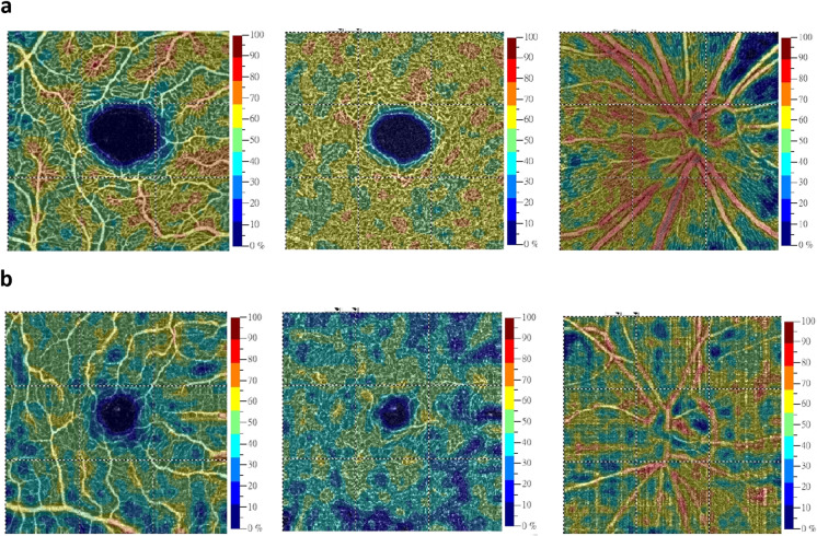
Fig. 3.
Representative optical coherence tomography angiography (OCT-A) scanning images in the macular and peripapillary regions from patients with different Hoehn-Yahr stages of PD. a The vessel density maps of the superficial retinal capillary plexus (left column), deep retinal capillary plexus (middle column), and radial peripapillary capillary layer in the peripapillary and disc regions (right column) in the right eye of a PD patient in Hoehn-Yahr stage I. b The vessel density maps of the superficial retinal capillary plexus (left column), deep retinal capillary plexus (middle column), and radial peripapillary capillary layer in the peripapillary and disc regions (right column) in the right eye of a PD patient in Hoehn-Yahr stage III

