Fig. 3.
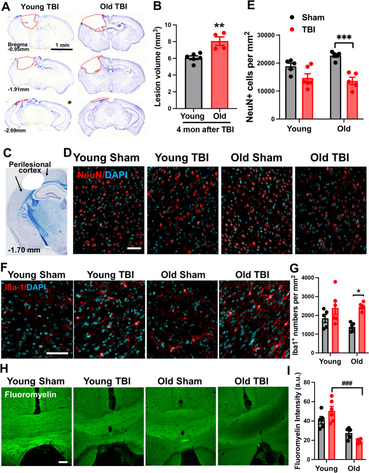
Aged mice exhibit exacerbated tissue damage, higher neuronal loss, and exaggerated white matter degradation after injury. A Representative images from lesion site of old TBI and young TBI mice at 16 weeks after injury. B Lesion volume was quantified based on the Cavalieri method of unbiased stereology using Stereologer 2000 software. N = 6 (young TBI) and 4 (old TBI). Unpaired t test, p < 0.01. C A diagram of a coronal section with arrows encompassing the perilesional cortex, adjacent to the impact site. D Neuronal count of NeuN-positive cells within the ipsilateral cortex showed significant neuronal loss in old TBI mice compared to old sham group. E Representative images of NeuN and DAPI. F–G Representative images and quantified Iba-1-positive cells. H–I Representative images and quantification data for FluoroMyelin staining in the medial corpus collosum. N = 5 (young sham), 5 (old sham), 6 (young TBI), and 5 (old TBI). Two-way ANOVA followed by Tukey’s post-hoc test. ***p < 0.001, *p < 0.05, vs old sham; ### p < 0.001, vs young TBI

