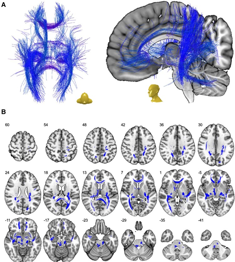
Figure 3.
Network of most affected white matter fibres. (A) Streamline-wise comparisons between COVID-19 patients and controls (with nuisance covariates ‘age’ and ‘sex’; exploratory threshold of P < 0.001) reveal a widespread network of white matter fibres in MNI space that were most affected by COVID-19-related V-CSF increase. (B) To display the distribution and extent of the aforementioned network in the white matter, 3D streamlines were rendered to generate a visiting map in MNI space (blue shading) that is superimposed onto a transversal T1-weighted template. Depicted in radiological orientation, i.e. left image side corresponds to patient’s right body side; numbers denote the axial (z) position in millimetres.

