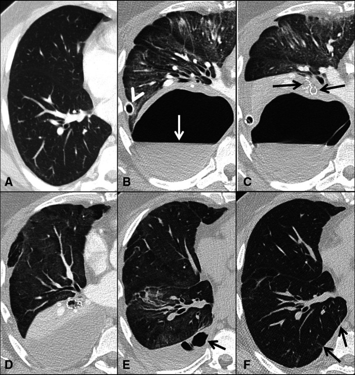
Figure 1.

(A–F). Serial transverse CT scans of the chest sections showing right hemithorax only. One year before admission for COVID-19 pneumonia routine CT with contrast agent was unremarkable except for some dependent atelectasis (A). CT pulmonary angiogram 3 weeks after admission shows a 10×18 cm cavitary lesion with an air-fluid level (arrow) and surrounding atelectasis of the lower lobe. A chest drainage tube is positioned in the periphery of the cavity (arrowhead). There are widespread ground glass opacities in the upper and middle lobe (B). Chest CT after insertion of endobronchial valves (arrows) covering all segments of the lower lobe (C). Three days later CT scans show endobronchial valves in situ and collapse of the lower lobe. The drainage tube is removed (D). One month later the endobronchial valves have been removed. In the lower lobe, there is almost complete re-expansion but still some opacifications related to COVID-19 pneumonia in all lobes. In the pleural cavity organised fluid with small air cavities (arrow) (E). High-resolution CT scans 5 months after admission show almost complete absorption of the abnormalities; however, small parenchymal bands are observed (arrows) (F).
