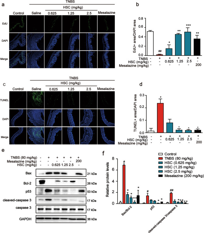
Fig. 3. The protective effect of HSC on intestinal epithelial cells.
a Sections of colon tissue were immunostained with EdU and observed by confocal laser-scanning microscope (n = 3). b The area ratio of EdU fluorescence staining intensity to DAPI. c Sections of colon tissue were immunostained with TUNEL and observed by confocal laser-scanning microscope (n = 3). d The area ratio of TUNEL fluorescence staining intensity to DAPI. e Western blotting analysis of the levels of Bax, Bcl-2, p53, and cleaved-caspase 3. Each experiment was repeated three times. f Statistical analysis of p53, Bax, Bcl-2, and cleaved-caspase 3 protein level. #P < 0.05, ##P < 0.01 vs control group; *P < 0.05, **P < 0.01, ***P < 0.001 vs TNBS group.

