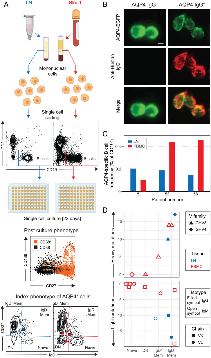
Fig. 3.
AQP4-specific B cells characterized from LNs and blood of patients with NMOSD. (A) Single B cells (CD3−CD19+) from paired blood (red) and LN (blue) samples were labeled with detection antibodies, index sorted as single cells, and cultured. By day 22, ∼50% of cells proliferate and differentiate into antibody-secreting cells (CD19 cells gated to show CD27, CD38, and CD138 expression). Indexing revealed the original B cell subsets: naive, double-negative (DN), IgD−, and IgD+ memory (Mem). (B) AQP4-reactive IgGs/IgMs in culture supernatants were identified by reactivity (red) directed against live HEK293T cells, which expressed surface AQP4–EGFP (green). (Scale bar, 10 µm.) (C) AQP4-specific B cell frequencies detected from three patients in LNs (blue) and PBMCs (red). (D) The heavy and light chains of these AQP4-specific BCRs (7 and 11 recovered, respectively) arose from all four studied B cell subsets and showed varied gene families (heavy variable IGHV3 as triangles and IGHV4 as diamonds) and light chain usage (kappa or lambda as squares or circles, respectively) from both LNs (blue) and PBMCs (red). The AQP4-reactive isotype detected in supernatants is shown using open (AQP4-IgM) or filled (AQP4-IgG) symbols; DN, double-negative cells; IGHV, immunoglobulin heavy chain variable region.

