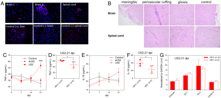
Fig. 5.
HEV invades the CNS tissues in HEV-infected pigs. Four-week-old HEV-negative SPF pigs were intravenously inoculated with the quasi-enveloped eUS2 virus or nonenveloped US2 or medium as control. Samples of brain and spinal cord were collected at 21 dpi. (A) The formalin-fixed tissues of brains and spinal cords were paraffin-embedded and in situ-hybridized with a fluorescent-labeled (red) HEV-specific probe (representative pictures showing the detection of HEV RNAs in CNS tissues by FISH are shown). (B) The tissues of brains and spinal cords were H&E-stained and paraffin-embedded. The histological lesions were evaluated, in a blind fashion, by a board-certified veterinary pathologist (T. L.). Representative histopathology pictures are shown. Weekly sera were collected from each infected pig and the serum levels of TNF-α (C) and IL-18 (E) were determined. Comparison of the serum levels of TNF-α (D) and IL-18 (F) between HEV-infected pigs with detectable HEV RNAs in brain tissues (n = 3) and infected pigs with no detectable HEV RNA in brain tissues (n = 4) at 21 dpi. (G) Total RNAs were isolated from the brain tissues of the US2-infected pigs at 21 dpi. The mRNA levels of the TJ proteins in brain tissues (occludin, ZO-1, VE-C, and caudin-5) were quantified by qRT-PCR. *P < 0.05, ****P < 0.0001, one-way ANOVA.

