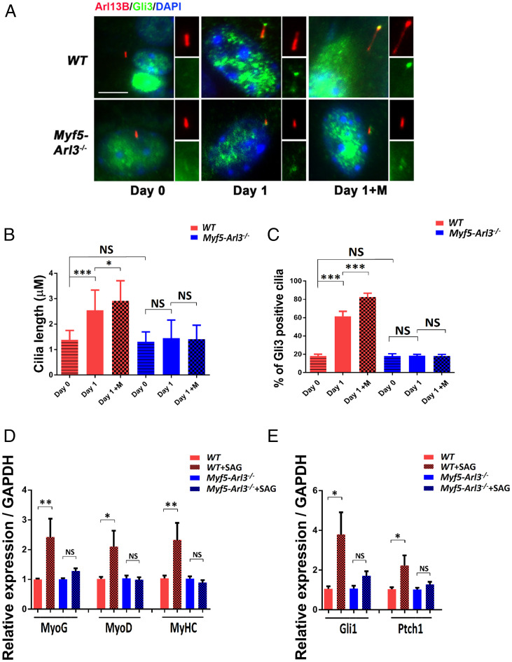
Fig. 6.
The activation of the Hh signaling pathway by mechanical stimulation depends on functional primary cilia. (A) GLI3 translocates to cilia after mechanical stimulation in WT but not Myf5-Arl3−/− myoblasts. Immunofluorescence staining of cilia with ARL13B (red) and GLI3 (green) and counterstained with DAPI (blue). GLI3+ cilia were counted. (Scale bar, 5 μm.) (B) The length of cilia was measured using a Nikon ECLIPSE Ti with MetaMorph software. An average of 300 cilia was measured in each group. (C) Quantification of A: 300 cells were counted for each line (five lines for each genotype). (D) Relative expression of MyoG, MyoD, and MyHC in myoblasts derived from WT and Myf5-Arl3−/− mice after SAG treatment using real-time PCR. (E) Relative expression of Gli1 and Ptch1 expression in myoblasts derived from WT and Myf5-Arl3−/− mice after SAG treatment using real-time PCR. Note the abrogation of Hh signaling markers in Myf5-Arl3−/− mice after mechanical stimulation in vitro. (One-way ANOVA with Tukey’s multiple comparisons tests, *P < 0.05, **P < 0.01, ***P < 0.001; NS, not significant.)

