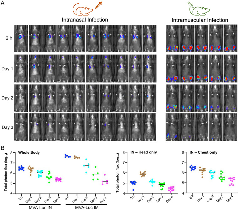
Fig. 3.
Live animal imaging of Luc following IN or IM inoculation of rMVA. (A) Mice (C57BL/6) were inoculated with 2 × 10⁷ PFU of MVA/Luc IN (n = 10) or IM (n = 5), the latter divided into each thigh. At the indicated hours and days, luciferin was injected intraperitoneally, and Luc was detected with a live animal imager. The exposure time and binning factor were kept constant. Ventral views of mice are shown. Intensity increases from blue to red. (B) Total photon flux (photons per cm2/sr) was determined for the whole body or head and chest separately. Data are representative of two independent experiments.

