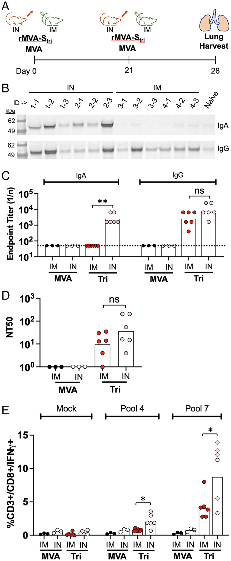
Fig. 4.
Immune responses in the lungs of vaccinated mice. (A) K18-hACE mice were vaccinated with 2 × 10⁷ PFU of rMVA-Stri IN (n = 6) or IM (n = 6) and then boosted 2 wk later via the same route. The mice were killed 1 wk after the boost and perfused lungs were excised, dissociated, and centrifuged to collect supernatant and cells. (B) Lung tissue supernatants of vaccinated mice and a naïve mouse were analyzed by SDS/PAGE and blots were probed with anti-IgA and anti-IgG antibodies. Positions of marker proteins in kilodaltons shown on left. (C) IgA and IgG SARS-CoV-2 S binding titers of the lung tissue supernatants were determined by ELISA. Values for individual mice and geometric mean are shown. The dotted line represents the limit of detection. (D) Pseudovirus neutralizing titers were determined on lung tissue supernatants. Values for individual mice and geometric mean titers are shown. (E) Lung cells were mock stimulated or stimulated with peptide pool 4 or 7 and the percent CD3+CD8+IFN-γ+ were determined by flow cytometry; 10,000 to 70,000 events were collected for each sample. Significance: *P ≤ 0.04, **P = 0.002; ns, not significant.

