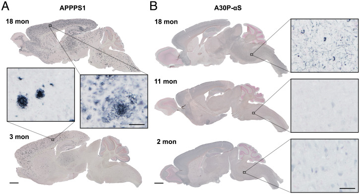
Fig. 1.
APPPS1 and A30P-αS mice show progressing proteopathic brain lesions. (A) Representative images of immunostained Aβ deposits in young (3-mo-old) and aged (18-mo-old) APPPS1 mice. Note the distinct Aβ plaques at 3 mo mainly in the neocortex and the appearance of Aβ deposition all over the brain at 18 mo of age. (B) Immunostained phosphorylated (pS129) αS in young (2-mo-old), adult (11-mo-old), and aged (end-stage disease; 18-mo-old) A30P-αS mice. (Scale bars, 1 mm for brain sections and 50 µm for Insets.) Counterstaining with nuclear fast red.

