Fig. 1.
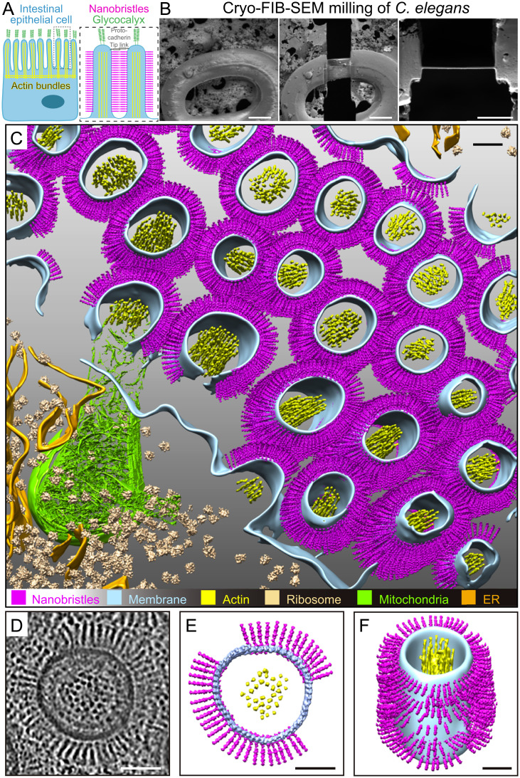
In situ cryo-ET of the C. elegans intestinal brush border reveals nanobristles on the lateral surface of microvilli. (A) A schematic diagram of an intestinal epithelial cell (Left) and two microvilli (Right) from the dotted box in Left. The glycocalyx and the protocadherin tip link are the characterized cell-surface structure at microvillar tips. This work shows that numerous nanobristles (magenta) decorate the lateral surface of microvilli. (B, Left and Center) Representative cryo-SEM images of the C. elegans L1 larvae before and after FIB milling. (Scale bars, 10 μm.) (B, Right) Representative FIB image of the ∼200-nm-thick cryo-lamella. (Scale bar, 5 μm.) (C) A 3D rendering of the C. elegans intestinal brush border showing various macromolecules and structures. Magenta, nanobristles; cyan, membrane; yellow, actin; beige, ribosome; green, mitochondria; orange, ER) Nanobristles and ribosomes were mapped back in the tomogram with the computed location and orientation. (D) A selected microvillus from E magnified for visualization. (E and F) Cryo-ET tomogram slices of microvilli (E, top view; F, side view). (Scale bars in C–F, 50 nm.)

