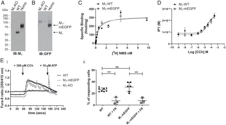
Fig. 4.
Expression and function of M1-mEGFP in neuronal cultures. Lysates from primary neuronal cells generated from day 16 embryos of WT, M1-mEGFP homozygous (Homo) knock-in and M1-knockout (KO) mice were resolved by SDS-PAGE and immunoblotted with either anti-M1 (A) or anti-GFP (B) antisera. (C) Specific binding of varying concentrations of [3H]NMS to intact neurons from such cultures of WT and homozygous M1-mEGFP–expressing mice was measured to allow assessment of both Bmax (maximum specific binding) and Kd for the ligand. Neither parameter was significantly different (P > 0.05) for the two sets of neurons. A representative example of three experiments is shown with error bars representing the SD (D). Such neuronal cultures from WT or homozygous M1-mEGFP–expressing mice were used to measure the production of inositol monophosphates in response to varying concentrations of carbachol (CCh). Cultures of combined hippocampal and cortical neurons from WT, homozygous M1-mEGFP–expressing and M1-KO mice maintained for 7 d were loaded with Fura-8-AM. (E, i) These were then imaged over time in the absence of or after exposure to 300 μM carbachol. In cells isolated from M1-KO mice, after exposure to carbachol, 10 μM adenosine triphosphate (ATP) was added to confirm that the cells were able to respond to an external stimulus. (E, ii) In certain experiments, cells were pretreated with the Gq/G11 inhibitor FR900359 (FR) (21). Data, shown as a percentage of cells tested that responded to carbachol are results taken from three to eight individual mice with analysis of between 31 and 93 cells from each animal. ***, P < 0.001. ns, not significantly different.

