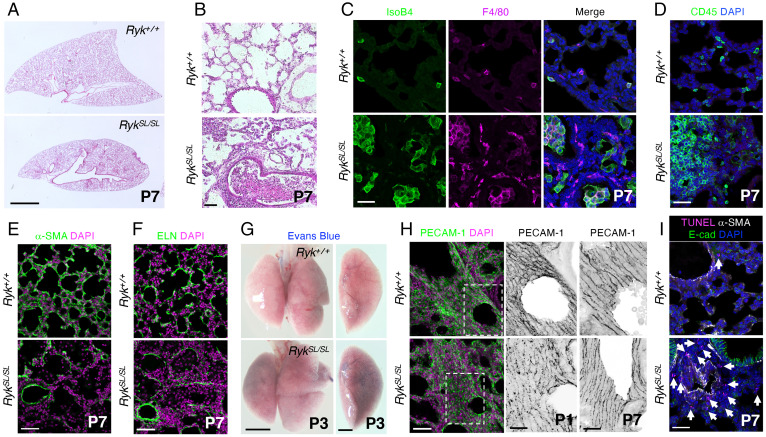
Fig. 1.
Ryk deficiency leads to lung hypoplasia and inflammation. (A) Hematoxylin and eosin (H&E) staining of P7 Ryk+/+ (n = 10) and RykSL/SL (n = 12) lung sections. (B) High magnification of H&E-stained P7 Ryk+/+ (n = 10) and RykSL/SL (n = 12) lung sections. (C) IsoB4 staining (immune cells) and F4/80 immunostaining (alveolar and interstitial macrophages) in P7 Ryk+/+ (n = 10) and RykSL/SL (n = 12) lung sections. (D) Immunostaining for CD45 (hematopoietic cells) in P7 Ryk+/+ (n = 10) and RykSL/SL (n = 12) lung sections. (E) Immunostaining for α-SMA (to mark myofibroblasts) in P7 Ryk+/+ (n = 10) and RykSL/SL (n = 12) lung sections. (F) Immunostaining for ELN (to mark secondary septa) in P7 Ryk+/+ (n = 10) and RykSL/SL (n = 12) lung sections. (G) Representative images of P3 Ryk+/+ (n = 6) and RykSL/SL (n = 6) lungs injected with Evans blue dye. (H) Immunostaining for PECAM-1 (endothelial cells) in P1 and P7 Ryk+/+ (n = 5) and RykSL/SL (n = 5) lung sections. High-magnification image of the areas in the dashed boxes is shown in the Middle panels. (I) TUNEL staining and immunostaining for α-SMA and E-cadherin in P7 Ryk+/+ (n = 10) and RykSL/SL (n = 12) lung sections. Arrows point to TUNEL-positive cells. (Scale bars: 2 mm for A and G, Left, 1 mm for G, Right; 50 μm for B, E, F, and H, Left; and 30 μm for C, D, H, Right, and I.)

