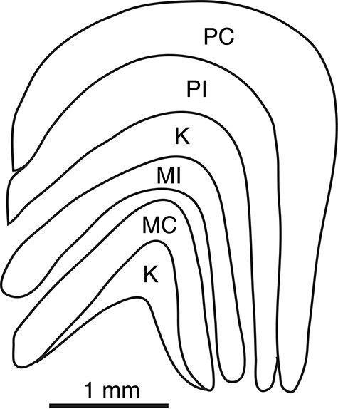
Figure 2 .

The lateral geniculate of a marmoset. The drawing is based on a coronal brain section with medial to the left. There are 4 obvious geniculate layers, including a pair of more dorsal parvocellular (M) layers and a more ventral pair of magnocellular (M) layers. The outer MC and PC layers receive retinal inputs from the contralateral retina, while the inner MI and PI layers receive inputs from the ipsilateral retina. Small koniocellular (K) neurons exist in the cell poor septal regions between layers, with a larger number between the inner M and P layers and below the MC layer. These septal regions of K neurons are not recognized as distinct layers.
