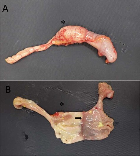
Figure 2 .

Duodenal dilation in the common marmoset. (A) The upper GI tract of a common marmoset with duodenal dilation. (B) The upper GI tract of the same marmoset with the tissue transected to expose the lumen. The duodenum (left) and stomach (right) are clearly demarcated (as denoted by the arrow) but are similar in diameter. The asterisk, in both images, is located at the dilated aspect of the duodenum. Images courtesy of Andres Mejia, WNPRC.
