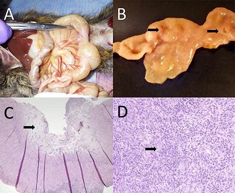
Figure 3 .

Gastrointestinal adenocarcinoma in the common marmoset. (A) Two strictures are present in the small intestine with associated discoloration and notable dilation proximal to each stricture. (B) The tissue has been transected and mucosal lesions (denoted by the two arrows) can be seen resulting in localized proliferation of the mucosa and strictures of the lumen. (C) Histology of gastrointestinal adenocarcinoma with mucosal erosion, edema and loss of normal mucosal architecture. The arrow denotes highly edematous tissue associated with the erosion. (D) Higher magnification photomicrograph of proliferative neoplastic cells effacing the intestinal mucosa. The arrow denotes a cluster of abnormal cells that are not forming normal glandular architecture within the tissue. Images courtesy of Heather Simmons and Andres Mejia, WNPRC.
