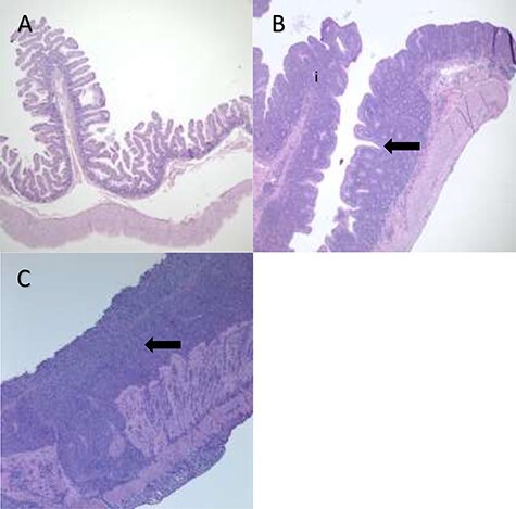
Figure 4 .

Degrees of Inflammation of the Small Intestine in the Common Marmoset. (A) Architecture of normal small intestines from a marmoset unaffected by CLE. Villi are thin and long, projecting into the lumen with few cells in the lamina propria. (B) Moderate small intestinal inflammation in a marmoset with CLE. The lamina propria is thicker with more inflammatory cells (i), and rounded, blunted villous projections (the arrow denotes an area in which there is little space between blunted villi). (C) Severe small intestinal inflammation in CLE with numerous inflammatory cells in the lamina propria and diffuse fusion of villi (denoted by the arrow). Images taken at 4X. Images courtesy of Heather Simmons and Andres Mejia, WNPRC.
