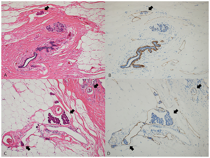
Figure 1.
Detection of low and high expression of LVI in H&E staining and D2-40 immunostaining specimens. A representative case with low LVI stained with (A) H&E and (B) D2-40. A representative case with high LVI stained with (C) H&E and (D) D2-40. Black arrows indicate LVI (all magnification, ×100). H&E, hematoxylin and eosin; LVI, lymphovascular invasion.

