. 2022 May 17;13(24):7080–7097. doi: 10.1039/d2sc01278b

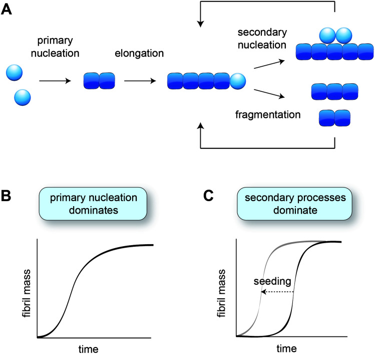
Fig. 1. Aggregation kinetics in vitro. (A) Schematic of the different microscopic aggregation steps. Spheres indicate monomeric protein, and cubes the monomeric subunits in the fibril. The nucleus is formed by two monomers in the case of amyloid-β, but may have a different size for other proteins. Secondary nucleation and fragmentation cause a positive feedback loop, leading to the generation of more fibrils. (B) Cartoon of an aggregation profile characterised by primary nucleation. (C) Cartoon of an aggregation profile characterised by secondary processes, evident from the long lag phase. Adding pre-formed fibril seeds shortens the lag phase by bypassing primary nucleation.

Fig. 1